Presentations made painless
- Get Premium

106 Ultrasound Essay Topic Ideas & Examples
Inside This Article
Ultrasound technology has revolutionized the field of medicine, allowing healthcare professionals to visualize internal structures and organs without invasive procedures. As a result, ultrasound has become an essential tool for diagnosing and monitoring various medical conditions. If you are a student studying ultrasound technology or a healthcare professional looking to expand your knowledge, here are 106 ultrasound essay topic ideas and examples to help you explore this fascinating field further.
- The history and development of ultrasound technology
- The physics behind ultrasound imaging
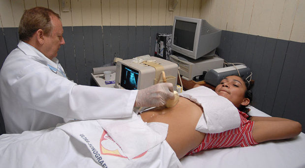
- The role of ultrasound in obstetrics and gynecology
- Ultrasound-guided procedures in interventional radiology
- The use of ultrasound in diagnosing musculoskeletal injuries
- Ultrasound imaging of the heart (echocardiography)
- The benefits and limitations of 3D/4D ultrasound imaging
- Contrast-enhanced ultrasound for liver imaging
- Ultrasound elastography for assessing tissue stiffness
- The role of ultrasound in diagnosing breast cancer
- Ultrasound imaging in emergency medicine
- Point-of-care ultrasound in critical care settings
- The use of ultrasound in vascular imaging
- Ultrasound-guided nerve blocks for pain management
- The future of ultrasound technology in healthcare
- Ultrasound imaging of the thyroid gland
- The use of ultrasound in diagnosing gallbladder disease
- Ultrasound-guided biopsy procedures
- Ultrasound imaging of the kidneys
- The role of ultrasound in diagnosing appendicitis
- Ultrasound imaging of the pancreas
- The use of ultrasound in diagnosing gastrointestinal disorders
- Ultrasound-guided injections for joint pain
- Ultrasound imaging of the urinary tract
- The benefits of portable ultrasound technology
- Ultrasound imaging of the prostate gland
- The use of ultrasound in diagnosing testicular conditions
- Ultrasound-guided drainage procedures
- Ultrasound imaging of the spleen
- The role of ultrasound in diagnosing hernias
- Ultrasound-guided nerve ablation for pain management
- Ultrasound imaging of the placenta
- The use of ultrasound in diagnosing fetal anomalies
- Ultrasound-guided thyroid biopsy procedures
- Ultrasound imaging of the adrenal glands
- The benefits of contrast-enhanced ultrasound for liver imaging
- Ultrasound-guided joint injections for arthritis
- Ultrasound imaging of the parathyroid glands
- The role of ultrasound in diagnosing lymph node abnormalities
- Ultrasound-guided breast biopsy procedures
- Ultrasound imaging of the thymus gland
- The use of ultrasound in diagnosing mediastinal masses
- Ultrasound-guided pleural procedures
- Ultrasound imaging of the pericardium
- The benefits of contrast-enhanced ultrasound for vascular imaging
- Ultrasound-guided nerve blocks for chronic pain management
- Ultrasound imaging of the carotid arteries
- The role of ultrasound in diagnosing peripheral vascular disease
- Ultrasound-guided varicose vein procedures
- Ultrasound imaging of the aorta
- The use of ultrasound in diagnosing deep vein thrombosis
- Ultrasound-guided sclerotherapy for spider veins
- Ultrasound imaging of the liver and biliary system
- The benefits of contrast-enhanced ultrasound for renal imaging
- Ultrasound-guided renal biopsy procedures
- The role of ultrasound in diagnosing adrenal tumors
- Ultrasound-guided adrenal vein sampling procedures
- Ultrasound imaging of the pancreas and spleen
- The use of ultrasound in diagnosing pancreatic cancer
- Ultrasound-guided pancreatic biopsy procedures
- Ultrasound imaging of the gallbladder and biliary system
- The benefits of contrast-enhanced ultrasound for pancreatic imaging
- Ultrasound-guided percutaneous cholecystostomy procedures
- Ultrasound imaging of the gastrointestinal tract
- The role of ultrasound in diagnosing inflammatory bowel disease
- Ultrasound-guided intestinal biopsy procedures
- Ultrasound imaging of the kidneys and urinary tract
- The use of ultrasound in diagnosing kidney stones
- Ultrasound-guided percutaneous nephrolithotomy procedures
- Ultrasound imaging of the female reproductive system
- The benefits of contrast-enhanced ultrasound for gynecologic imaging
- Ultrasound-guided ovarian cyst aspiration procedures
- Ultrasound imaging of the male reproductive system
- The role of ultrasound in diagnosing testicular cancer
- Ultrasound-guided testicular biopsy procedures
- Ultrasound imaging of the musculoskeletal system
- The use of ultrasound in diagnosing sports injuries
- Ultrasound-guided joint aspiration procedures
- Ultrasound imaging of the nervous system
- The benefits of contrast-enhanced ultrasound for neuroimaging
- Ultrasound-guided nerve conduction studies
- Ultrasound imaging of the head and neck
- The role of ultrasound in diagnosing thyroid nodules
- Ultrasound-guided thyroid fine-needle aspiration biopsy procedures
- Ultrasound imaging of the chest and lungs
- The use of ultrasound in diagnosing pleural effusions
- Ultrasound-guided thoracentesis procedures
- Ultrasound imaging of the heart and blood vessels
- The benefits of contrast-enhanced ultrasound for cardiac imaging
- Ultrasound-guided cardiac catheterization procedures
- Ultrasound imaging of the liver and spleen
- The role of ultrasound in diagnosing liver cirrhosis
- Ultrasound-guided liver biopsy procedures
- Ultrasound imaging of the pancreas and biliary system
- The use of ultrasound in diagnosing pancreatic pseudocysts
- Ultrasound-guided percutaneous drainage procedures
- Ultrasound imaging of the gastrointestinal tract and kidneys
- The benefits of contrast-enhanced ultrasound for urologic and gastrointestinal imaging
- Ultrasound-guided percutaneous nephrostomy procedures
- Ultrasound imaging of the female reproductive system and bladder
- The role of ultrasound in diagnosing pelvic organ prolapse
- Ultrasound-guided bladder sling procedures
- Ultrasound imaging of the male reproductive system and prostate
- The use of ultrasound in diagnosing benign prostatic hyperplasia
- Ultrasound-guided prostate biopsy procedures
These essay topic ideas and examples cover a wide range of ultrasound applications and specialties, providing you with ample opportunities to explore and research this exciting field further. Whether you are a student or a healthcare professional, delving into these topics can deepen your understanding of ultrasound technology and its role in modern medicine.
Want to research companies faster?
Instantly access industry insights
Let PitchGrade do this for me
Leverage powerful AI research capabilities
We will create your text and designs for you. Sit back and relax while we do the work.
Explore More Content
- Privacy Policy
- Terms of Service
© 2024 Pitchgrade
On Medicine

The Future, High-Tech Impacts of Ultrasound in Health Care
Ultrasound technology is a vital part of health care. It is utilized for a variety of diagnostic purposes, but until recently, all an ultrasound could be used for was viewing the inside of the human body. New advances in this technology have shown great promise in internal medicine applications. Where can ultrasound be applied in internal medicine, and what implications does it have for the future of ultrasound technology?
Kayla Matthews 18 Apr 2017
Current Ultrasound Applications
Ultrasounds are traditionally used for imaging, usually in internal medicine and prenatal applications. Common newer applications for ultrasound technology include:
- Shock Wave Lithotripsy — This process utilizes targeted ultrasound waves to break up kidney stones and other calcium-based growths in the body. It’s most commonly used to break up stones that are too large to pass, or ones that will not pass naturally. It is an alternative to surgery.
- Dental Descaling — Ultrasounds have been used for periodontal therapy in the form of ultrasound debridement. When used correctly, ultrasound debridement is more efficient and effective than manual debridement.
There are other less common uses for ultrasound technology, but they are not as widely applied or studied.
- Ultrasound for Liver Tumors
Traditional treatment for liver cancer and tumors is to surgically remove the malignant tissue, then treat any remaining cells with a combination of radiation and chemotherapy. By utilizing targeted ultrasound waves, liver tumors can be destroyed without the need for surgical intervention. The waves, emitted by more than 1,000 micro-emitters, cauterize the tissue inside the body.
It is not an easy procedure — the liver moves with the patient’s breath, so treatment requires the patient to either hold their breath or be sedated. The liver also lies behind the rib cage, so the emitters need to be calibrated to avoid doing damage to the ribs. By pairing real-time MRI imagery with these micro-emitters, surgery can be avoided by destroying the malignant cells without the need for invasive surgery.
Additionally, the use of ultrasound technology to cauterize tumors internally would prevent the creation of surgical smoke, which can cause a dangerous decrease in surgeon visibility . Surgical smoke is a hazard for any procedure that utilizes electro-cauterization techniques.
- Immunotherapy-Based Cancer Treatment
Immunotherapy treatment for cancer has become a companion for traditional treatments, utilizing medication to stimulate the patient’s own immune system to fight cancer cells. Many of these new treatments have been approved by the FDA, but they are just starting to become commonplace.
A new treatment technique, currently in preclinical trials , pairs these immunotherapy treatments with ultrasound technology. Early findings suggest ultrasound treatment helps improve a patient’s immune response, helping their body’s immune system fight cancer more effectively. The treatment is noninvasive and unlike radiation therapy, it doesn’t have any negative side effects. It also doesn’t negatively affect the immune system.
- Bone Healing and Joint Therapy
Broken bones, traditionally, take anywhere from 3 to 10 weeks to heal, depending on the age and health of the patient and the severity of the break.
By utilizing low-intensity pulsed ultrasound (LIPUS), studies have shown increased healing rates of anywhere from 24 percent to 42 percent when applied to fresh fractures. While healing rates do drop for older fractures, overall success rates are high. Some success rates are upwards of 80 percent.
The exact reason ultrasounds help bones knit is unknown, but it has seen a unique application in the form of a 3-D printed cast that aids healing with the use of ultrasound waves. While it’s only a prototype, it could easily become the next advancement in bone repair.
Ultrasound technology could potentially change the way noninvasive surgery and treatments are done. These advances can save lives, decrease healing time and do a whole lot more than just look inside the body.
View the latest posts on the On Medicine homepage
- Latest Posts

Kayla Matthews
Latest posts by kayla matthews ( see all ).
- The Future, High-Tech Impacts of Ultrasound in Health Care - 18th April 2017
- US traffic fatalities and relationship to medical cannabis laws - 10th February 2017
Recommended posts
Popular on medicine tags.
- BMC Medicine
- clinical trials
- medical evidence
- Genome Medicine
- ISRCTN Registry
- Alzheimer's
- Alzheimer's Research & Therapy
- breast cancer
Popular posts
- Most shared
Sorry. No data so far.
Most Shared Posts
- Photobiomodulation therapy to prevent chemotherapy-induced oral mucositis
- A unique approach. A unique population. Investing in nurses’ mental health
- Does the midwife-led continuity of carer model improve birth outcomes and maternal mental health in vulnerable women?
- A proactive diabetes review model: from concept to nurse-led research
- Clinical Trials Day 2023: what about the trial participant?
- September 2024 (1)
- March 2024 (1)
- February 2024 (2)
- January 2024 (1)
- November 2023 (1)
- October 2023 (1)
- September 2023 (7)
- July 2023 (1)
- June 2023 (1)
- May 2023 (4)
- April 2023 (1)
- March 2023 (2)
Home — Essay Samples — Nursing & Health — Nursing — The Rising Demand for Ultrasound Technicians
The Rising Demand for Ultrasound Technicians
- Categories: Medical Ethics Nursing
About this sample

Words: 637 |
Published: Mar 16, 2024
Words: 637 | Page: 1 | 4 min read
Table of contents
Educational requirements, job responsibilities, salary potential, future outlook.

Cite this Essay
Let us write you an essay from scratch
- 450+ experts on 30 subjects ready to help
- Custom essay delivered in as few as 3 hours
Get high-quality help

Dr Jacklynne
Verified writer
- Expert in: Nursing & Health

+ 120 experts online
By clicking “Check Writers’ Offers”, you agree to our terms of service and privacy policy . We’ll occasionally send you promo and account related email
No need to pay just yet!
Related Essays
2 pages / 777 words
3 pages / 1394 words
1 pages / 630 words
2 pages / 800 words
Remember! This is just a sample.
You can get your custom paper by one of our expert writers.
121 writers online
Still can’t find what you need?
Browse our vast selection of original essay samples, each expertly formatted and styled
Related Essays on Nursing
Nursing leadership is an essential component of healthcare delivery, with effective leadership playing a critical role in providing quality patient care and improving healthcare outcomes. This essay will explore the definition, [...]
American Nurses Association. (2021). Nursing: Scope and Standards of Practice (4th ed.). Silver Spring, MD: American Nurses Association.Nightingale, F. (1860). Notes on Nursing: What It Is, and What It Is Not. Harrison and [...]
A good nurse possesses a unique blend of qualities that extend far beyond medical knowledge and technical skills. In this essay, we will explore the essential qualities of a good nurse and how they significantly contribute to [...]
Clinical systems play a crucial role in modern healthcare delivery, influencing efficiencies and outcomes within nursing practice. This essay aims to explore the impact of clinical systems on healthcare delivery and nursing [...]
Bureau of Labor Statistics. ( 2019, September 4). Registered nurses. Retrieved from
In the realm of healthcare, integrity is a pillar that upholds the trust between patients and healthcare professionals. Nowhere is this more evident than in the field of nursing, where integrity serves as the bedrock of ethical [...]
Related Topics
By clicking “Send”, you agree to our Terms of service and Privacy statement . We will occasionally send you account related emails.
Where do you want us to send this sample?
By clicking “Continue”, you agree to our terms of service and privacy policy.
Be careful. This essay is not unique
This essay was donated by a student and is likely to have been used and submitted before
Download this Sample
Free samples may contain mistakes and not unique parts
Sorry, we could not paraphrase this essay. Our professional writers can rewrite it and get you a unique paper.
Please check your inbox.
We can write you a custom essay that will follow your exact instructions and meet the deadlines. Let's fix your grades together!
Get Your Personalized Essay in 3 Hours or Less!
We use cookies to personalyze your web-site experience. By continuing we’ll assume you board with our cookie policy .
- Instructions Followed To The Letter
- Deadlines Met At Every Stage
- Unique And Plagiarism Free
Ultrasound Technology in Podiatry Surgery Research Paper
- To find inspiration for your paper and overcome writer’s block
- As a source of information (ensure proper referencing)
- As a template for you assignment
Introduction
Randomized controlled trial, educational intervention, theoretical/conceptual framework, literature review strategy, research design, chapter analysis, research questions and hypotheses, scope, limitations, and delimitations.
Technology has already become a part of people’s lives in many spheres. The Healthcare system heavily relies on various technological advances as they provide manifold opportunities to improve the services available to patients. Technology helps in treating illnesses and managing individuals’ health conditions. It is also associated with efficiency, error-free procedures, faster healing processes. Podiatry surgery also benefits from the utilization of technology.
One of the areas where technology has proved to be essential is minimally invasive surgery. For instance, endoscopy has become a widely used procedure that minimizes the size of incisions and leads to a faster and less painful healing process (De Leeuw, Van Sterkenburg, Van Bergen & Van Dijk, 2011). The risk of infection is also reduced. Another important technological advancement is associated with the external fixation that is now performed with the use of materials that enable minimum incisions and intrusion into the blood circulation (Didomenico, Ziran & Cane, 2011).
However, ultrasound surgery and color Doppler-guided surgery are now seen as some of the most recent advances that maximize the efficiency of various procedures. For instance, Alfredson and Isaksson (2014) note that these methods are effective when it comes to “chronic painful insertional Achilles tendinopathy” (p. 7). The healing process is less painful and quite shorter. The patients who took part in the study reported that they were satisfied with the outcomes of the treatment. Therefore, it is clear that various technological advances enable surgeons to carry out various procedures in a more effective way.
It has been acknowledged that a randomized controlled trial (RCT) provides sound evidence to support or refute hypotheses in clinical research. At that, Bowen et al. (2012) note that RCT should be characterized by “sufficient detail for replication” to be used as the ground of any clinical research (p. 4). First, it is important to briefly outline the peculiarities of the RCT to understand the researchers’ point.
Randomized controlled trials involve participants who are randomly chosen, which eliminated any bias related to sampling. People of different backgrounds with different health histories often take part in the research, which allows researchers to understand the effectiveness of treatment (procedure, medication, and so on) in a wide population (Gordon, Darbyshire & Baker, 2012). It is possible to note that this is one of the most reliable tools to implement research. However, the lack of details when reporting the results of a trial can undermine the relevance of a study. If the trial cannot be replicated, the results may seem unreliable as there is no way to check if all the procedures were followed properly and whether all results were interpreted carefully.
Gordon et al. (2012) stress that the provision of sufficient details enables researchers to check the validity of the RCT and come to sound conclusions concerning the issue under study. Thus, it is crucial to make sure that the trial is properly reported and can be easily replicated to encourage other researchers to check the results of the trials implemented. Peer reviews are major tools to contribute to the knowledge base and come up with important insights into a variety of healthcare issues and concerns.
Intervention
The proposed educational intervention is associated with the use of ultrasound in podiatry surgery. The use of ultrasound is instrumental in identifying the exact areas of concerns that eliminates possible errors as well as minimizes the incisions. The educational intervention includes reading and discussion of several articles concerning the use of the technology mentioned.
The study by Alfredson and Isaksson (2014) can be central to the intervention. Learners can even try to replicate this study (in particular, the part concerning examination). Several videos and visuals are shown during the discussion sessions. The next stage is the learners’ review of the existing literature. The discussion of the information learned is the following stage.
The practice is the final stage of the intervention. Learners practice using the tool and evaluating the results of the examination. The assessment will include the focus on the ability to use the technology as well as learners’ willingness and readiness to use it in their clinical practice. The learners will identify the affected tissue and health conditions following the examination results. They will also carry out the examination. Finally, they will complete a survey concerning their views on technology and their willingness or readiness to use it in their clinical experiences.
Assumptions
The learners will be able to use ultrasound in podiatry surgery effectively. They will be able to use the technology in numerous settings. They will also be willing to try new ways of employing the tool by implementing their trials. Besides, the participants will feel empowered to initiate acquiring tools and technology to improve their performance as well as the performance of the healthcare facility.
Limitations
One of the major limitations is the fact that the intervention will cover a limited number of learners. Furthermore, specific equipment (characterized by certain features) will be used, which limits the learners’ ability to use other types of equipment. The duration of the intervention will be quite brief. However, the intervention can be extended if necessary. It is necessary to add that the evaluation of the intervention will help identify the most appropriate duration of the training course.
Delimitations
The intervention will last for two weeks. The learners will have four training sessions a week and one assessment session during the second week. Ten novice surgeons will participate. This sample is chosen as novice surgeons may lack the necessary knowledge and skills necessary to carry out certain operations with the use of ultrasound technology.
The intervention will remove such gaps if any. The equipment available at the healthcare facility will be employed. The evaluation of the intervention effectiveness will be held within the period of one and three months after its beginning. The extent to which the technology is utilized, patients’ satisfaction, the quality of services provided will be used as criteria for the assessment of the training.
Operationalization of Terms
Several terms will be employed in the study. Ultrasound technology is the equipment used in a healthcare facility to examine the Achilles insertion. Novice surgeons are surgeons who have worked from 3 to 9 months in the healthcare facility.
Theoretical Framework
When conducting research, it is critical to employ an appropriate theoretical framework. The choice of the framework depends on the focus of the study, the field of study, and so on. When it comes to medical technology, it is possible to use several theories. One of these frameworks can be a motivation theory (Geisler & Heller, 2012). It is possible to employ Alderfer’s ERG theory as it helps unveil the motivation of healthcare professionals to utilize technology. Thus, existence, relatedness, and growth need to drive people’s behaviors. When using technology, surgeons will be able to satisfy their growth needs.
Other motivation theories can also be used to identify the extent to which healthcare professionals are committed to using this or that technological advancement. Maslow’s hierarchy of needs can help evaluate the commitment of the medical staff as regards their physiological, safety, belongingness, esteem, and self-actualization needs. It is also possible to use Maslow’s theory of the hierarchy of needs to explore patients’ views concerning the use of medical technology in their treatment. This study can explore the way patients see the benefits and downsides of the use of technology. Their fears and concerns can also be examined. Therefore, the study may detect barriers to the use of technology and ways to overcome them by using methods that will satisfy patients’ needs.
As far as the use of medical technology is concerned, it is possible to employ the utility theory as well. The utility theory focuses on the way a product or service can satisfy the needs of the consumer who chooses following several criteria (Geisler & Heller, 2012). The use of technology is not confined to purely clinical outcomes. Healthcare facilities have quite limited funds and management has to decide which technology to use, and the choice can be rather difficult due to the abundance of products available. The utility theory framework can help identify the most appropriate tools and equipment for a particular health care facility.
Another theoretical approach is associated with professional dominance theory. According to this theory, healthcare professionals seek control over choices made within an organization (Cockerham, 2015). The theory was widely used in the 1970s, but it is still applicable and can be utilized to explore the way technology is chosen and employed in healthcare facilities. The focus can be on the way doctors affect the choices and shape the technological equipping of hospitals. It is also possible to use this framework when evaluating the effectiveness of training associated with the use of technology. The theory can help the researcher identify the extent to which healthcare professionals feel empowered and initiate the use (as well as the purchase of some equipment).
Conceptual Framework
As to the conceptual framework, it is possible to employ several concepts. The first concept to be used is knowledge (Geisler & Heller, 2012). Technology is always connected with the concept of knowledge and the creation of technology as well as its proper usage in the clinical setting involves a certain degree of knowledge and skills. Knowledge management and sharing are also closely connected with medical technology. Therefore, knowledge is a key element of the use of technological advances. Training is closely connected with the concept of knowledge. The medical staff needs training as technological advances appear each day, and healthcare professionals should have the necessary training to utilize new equipment effectively.
Another important concept guiding the research associated with medical technology is administration (Geisler & Heller, 2012). The choice of technology to be purchased often depends on the facility’s management. Therefore, the concept of administration is also very important when focusing on medical technology. Administrators are often free to allocate funds to purchase equipment and invest in staff training. More so, the way the healthcare facility is equipped depends on the views of the administrators’ views on the matter. If the administrator does not find technology essential, the healthcare facility may lack the necessary resources.
Applicability is another concept to be considered. As has been mentioned above, many healthcare facilities have limited resources and have to prioritize. The choice of the most appropriate and relevant equipment is critical. It is essential to have the technology that will be used to its full potential. It is also important to make sure that the equipment will help address the most urgent issues the healthcare facility deals with. This can require the implementation of the research concerning the most common health issues patients have in this or that community.
Finally, it is also possible to employ the concept of motivation. In many cases, people tend to use something they know well (Geisler & Heller, 2012). Many people are reluctant to use innovations due to their fear of errors. Therefore, healthcare professionals should be motivated to use technological advances. They should try to use new tools and methods in various settings to achieve high results and provide high-quality healthcare services.
When implementing any research, it is critical to implement a comprehensive literature review. It provides the background for the research as the research obtains the information on the existing knowledge on the matter as well as existing gaps (Ridley, 2012). It can also help the researcher to choose the most appropriate methodology to implement the research.
Many approaches to search appropriate sources to review exist. Ridley (2012) identifies five broad categories that include catalogs, Internet search engines, bibliographical databases, open-access databases, and professional organization websites. As for the catalogs, it is possible to use the Library of Congress catalog to track sources available in US libraries. It is also possible to use BUBL Link to trace online resources (Ridley, 2012).
As far as databases are concerned, it is possible to use EBSCO databases as this online platform contains links to a wide variety of academic sources in numerous fields including medical technology and surgery. Medline is another helpful database that can provide the researcher with links to relevant sources. Of course, PubMed is also a valuable source that can be used to search relevant academic resources on health care issues and medical technology.
One of the most effective search engines is Google Scholar. It provides links to various resources including books, peer-reviewed articles, relevant magazines, and websites. Importantly, all these sources are associated with advanced search tools that help the researcher to refine the search of resources following keywords, topics, publishing dates, and so on. All these features save the researcher’s time and make the process of data collection for the literature review quite fast and efficient.
When implementing the search for resources, it is important to choose the relevant keywords to make the process fast and efficient. The keywords for this study can be as follows: podiatry surgery, medical technology, ultrasound technology, technological advances. These keywords will be instrumental in allocating relevant sources.
Quantitaive Research Methodology
Education is one of the pillars of the development of society. It is also a basis of the healthcare system as education is the platform for the transition of knowledge and skills from seasoned professionals to novice practitioners. Education within healthcare bears some traits of education in other spheres, but it also has certain peculiarities.
Moriates, Dohan, Spetz, and Sawaya (2015) stress that education within healthcare should provide professionals who have a set of competencies that enable them to provide high-quality healthcare service. Importantly, these competencies include particular clinical skills, knowledge of policies and financial issues (costs, insurance), as well as emotional intelligence, leadership, the use of technology, mentoring, advocacy, and so on (Moriates et al., 2015). One of the most distinctive features of healthcare education is its possible outcomes as healthcare professionals mainly deal with people’s health. No errors or even gap can be tolerated in this sphere.
The education within healthcare is similar to education in other fields, as apart from acquiring knowledge and skills to provide care, healthcare professionals learn to be an effective researcher to expand the knowledge base and develop new methods and tools to improve the services provided or the entire system. Furthermore, education is not confined to formal methods that involve studying in medical schools.
On-job training is an important component of the system that enables novel, as well as experienced, healthcare professionals to acquire new knowledge and skills to be able to provide high-quality services (Lochmiller & Lester, 2015). This is especially true when the use of technology is involved since technology is constantly changing and upgrading, which makes it important for practitioners to have the necessary skills to use it.
As far as the on-job training, the constructivist theory is an appropriate theoretical paradigm as it is consistent with the peculiarities of this type of education. The theoretical model implies the focus on people’s previous experience as well as collaboration (Pritchard & Woollard, 2013). This theory is applicable as employees tend to use their background knowledge to construct new knowledge and skills, and this process is facilitated through sharing ideas and experiences (Pritchard & Woollard, 2013). Thus, on-job training presupposes that employees already have certain knowledge that is expanded through learning new information and developing certain competencies.
The focus of this study is the examination of the effectiveness of an intervention aimed at improving surgeons’ competence to use ultrasound and color Doppler-guided technology to treat patients suffering from insertional Achilles tendinopathy. Alfredson and Isaksson (2014) report the positive impact of the use of this technology in surgeons’ practice. The intervention will cover novice surgeons. To evaluate the efficiency of the intervention, it is possible to employ a quantitative research design.
The quantitative design is associated with the analysis of numerical data, which, in turn, is characterized by a high degree of generalizability of findings. Thus, the quantitative analysis will allow the researcher to identify whether the intervention can be effective on a large scale. In other words, this type of research design will reveal the outcomes of intervention as an experiment will be carried out. When compared to the qualitative methodology, quantitative studies are regarded as more measurable and generalizable as variables that can be quantified are used. Within the quantitative research methodology, it is possible to consider two methods: a randomized controlled trial and quasi-experimental research (Greener, 2011).
First, it is important to note that these tools are very similar but differ in one of the key points. Both methodologies usually imply the use of an experiment. Thus, two groups participate, and one group receives certain intervention while the other does not get any training. The test group has the training intervention while practitioners in the other group receive some manuals with a list of properties and features, as well as for instructions, concerning the use of the new technology. The performance of the participants is measured with the use of such criteria as the frequency of the use of the technology, occurrence of errors, patients’ satisfaction.
O’Dwyer and Bernauer (2014) state that the major difference between quasi-experimental and true experimental (randomized controlled trial) designs is concerned with sampling. In the randomized controlled trial, the researcher randomly assigns people to the two groups (O’Dwyer & Bernauer, 2014). In a quasi-experimental study, the researcher chooses participants without randomization, which can be explained by the unavailability of participants (for instance, a specific group is in the researcher’s lens).
At that, when choosing a methodology, it is crucial to remember that the randomized controlled trial is associated with a greater degree of relevance as it is more generalizable. Any person has a chance to enter any type of group, which makes the evaluation more effective as the researcher can potentially estimate the way an average practitioner can benefit from the intervention.
In this study, it is possible to employ a randomized controlled trial as the researcher can randomly choose participants. The number of practitioners characterized by the dependent variable (the time they work for this healthcare facility) is sufficient, and several surgeons can receive the intervention while the rest can be provided with the manuals.
Importantly, other variables will be excluded, which means that the intervention’s effectiveness will be checked with a potentially wide population. In its turn, this will contribute to the generalizability of the findings. The researcher will identify the way an average practitioner can benefit from the intervention in question. On the contrary, if a specific group of people will participate in the training program, it will be unclear whether other practitioners will equally benefit from the intervention.
In conclusion, it is possible to note that education within healthcare, like the one in any other field, aims at transferring knowledge to the new generation. On-job training is critical for the healthcare system especially when it comes to the use of technology as it constantly evolves. When considering such training, it is possible to apply the constructivist learning theory as it implies the use of people’s background knowledge and sharing ideas. The collaboration is a key element of this approach, and the intervention in question is also characterized by a significant emphasis on collaboration.
It is necessary to note that research is an important component of education as well. An intervention aimed at the development of skills necessary to use ultrasound technology can be evaluated with the help of a quantitative study. It is possible to use randomized controlled trial or quasi-experimental research, but the former is preferable as it is associated with a greater degree of generalizability. In the randomized controlled trial, sampling procedures allow the researcher to generalize data, which is important for the evaluation of any training program.
Quasi-Experimental Design
This type of research design focuses on a non-random assignment. As a rule, a quasi-experimental design requires a provision of both pretest and posttest procedures of the two groups under comparison. The core of this design is causal hypotheses that need to be either proved or refuted.
Speaking of the conditions, it is essential to pinpoint that treatment versus no treatment (comparison) groups are usually compared. Among relevant techniques to evaluate the results of the study, scholars note “regression discontinuity design (RDD) and propensity score matching (PSM)” (White & Sabarwal, 2014). The above tools constitute the two principal algorithms for the quasi-experimental design.
However, a statistical analysis here is complicated by the very fact of the lack of randomization. In particular, Handley, Schillinger, and Shiboski (2011) emphasize that it creates possible non-equivalence between studied groups. Therefore, it seems beneficial to consider a randomized controlled trial design to compare it with the already discussed quasi-experimental design and choose the most appropriate way to conduct the research.
In its turn, the randomized control trial (RCT) offers greater relevance by definition. Its sampling assumes random assignment of participants to the two groups (O’Dwyer & Bernauer, 2014). Randomization is crucial in conducting this method of design. It should provide a random distribution of patients that is not dependent on any factors and comparability of the groups being compared by clinical and demographic characteristics of patients including the severity of the underlying disease under study, concomitant pathology, and therapy. Therefore, the randomized controlled trial is regarded as the most scientifically rigorous method of hypotheses assessment.
Rationale for Choosing the most Appropriate Design
As it was stated in previous sections, the purpose of the research is to study the effectiveness of an intervention focused on enhancing surgeons’ proficiency to utilize ultrasound and color Doppler-guided technology in patients with insertional Achilles tendinopathy. In this connection, the use of the randomized controlled trial is much more applicable due to its high generalizability. In other words, a wide population might be embraced by the researcher to make the study more credible. Jin, Hua, and Cao (2016) also note an increased evidence-based relevance and popularity of the randomized controlled trials in a clinical environment. The results of the study would contribute to the experience of average practitioners.
Data Collection and Evaluation Instruments
To conduct a quantitative study that was chosen before, it is essential to use the survey data collection. According to Cleophas (2012), survey questions allow data monitoring and acquiring that is crucial to achieving the established research goals. In the context of the quantitative study, a statistical analysis would serve as the most opportune method of data examination. The focal objective of the statistical analysis of the RCT is the establishment of a difference and the degree of its credibility concerning outcomes between the group with the test intervention and the control group.
Currently, there are plenty of packages of programs for statistic analysis of the results such as BMDP, SOLO, and others. However, to address the research questions, it is useful to apply SurveyMonkey data collection mechanism and subsequent SPSS analysis.
Antonius (2012) states that this is a significant instrument to analyze data effectively and accurately. Moreover, to obtain objective information on the effectiveness of the intervention, the analysis should include all originally randomized patients (intention-to-treat analysis) and those whose treatment was carried out in strict compliance with the study protocol (on protocol analysis) (Antonius, 2012). This is one of the key ways to minimize possible errors when the intention-to-treat analysis is based on the assumption that all patients received the treatment prescribed at randomization.
The previous chapters provide an in-depth analysis of the research designs and methods. First, it was revealed that the chosen topic of digital intervention in invasive podiatry surgery is of great importance in a modern healthcare system. In particular, the impact of ultrasound and color Doppler-guided technology was chosen to examine in detail. After that, several educational intervention peculiarities including assumptions (effectiveness of technology implementation), limitations (limited number of learners and specific equipment), delimitations (two weeks and four training sessions), and others were identified.
Furthermore, both theoretical and conceptual frameworks were delineated. The first one comprised such theories as motivational, Alderfer’s ERG, utility, and professional dominance while the second one employed the following concepts: knowledge, administration, and applicability. Databases search was chosen as the paramount literature review strategy, yet it would require the use of relevant keywords that might be as follows: podiatry surgery, ultrasound technology, or technological advances.
Speaking of the methodology, a direct connection between the purpose statement and research questions was detected. Alignment of the four fundamental elements including research method, design, purpose statement, and research questions was found crucial for the effectiveness of the study as they serve in the role of a basis. Finally, the last section examined both quasi-experimental and randomized controlled trial designs pointing out the evident advantages of the latter for this research. Also, it was stated that the quantitative study analysis in the framework of SPSS would be appropriate to present credible results.
Quantitative Dissertations Review
Medical research can be implemented with the use of qualitative and quantitative tools. It has been acknowledged that quantitative studies are characterized by a significant degree of generalizability which allows practitioners to apply the findings in various settings (Koop, 2011). It is necessary to note that studies based on the use of the quantitative research design may involve a research question (or several research questions), hypothesis (or hypotheses) or both. In many cases, both hypotheses and research questions are available. It is possible to review several quantitative dissertations to understand the peculiarities of effective research questions and hypotheses.
As far as the hypothesis is concerned, it can be referred to as the major expectation of the researcher. It should also be as detailed as possible to enable the researcher to see whether the goals of the study have been met (Lum, 2008). It is essential to make sure that the hypothesis is consistent with the purpose of the study (Koop, 2011). There are different approaches to crafting hypotheses. For example, Johnson (2008) provides a null hypothesis that is proved to be true.
When it comes to research questions, they can be defined as particular objectives of the study or specific elements of the issue under analysis. Eshun and Eshun (2013) put special questions or the so-called Wh- questions. This provides a certain plan for evaluation of the intervention under analysis. These questions should also be very detailed and consistent with the hypothesis (or hypotheses) identified (Johnson, 2011; McLaughlin, 2012).
The Research Questions and Hypothesis for the Present Study
This study aims at evaluating the effectiveness of an intervention. The researcher is specifically interested in such areas as the frequency of the use of the equipment, peculiarities (settings) of this use, patients’ satisfaction and the participants’ commitment to contribute to the development of the healthcare facility through the improvement of technology-associated policies. The research questions of the present study can be formulated as follows:
- How does the intervention correlate with the frequency of the use of technology, patients’ satisfaction, as well as the quality of services provided?
- How does the intervention affect the participants’ willingness to implement their research or find new ways of utilizing the equipment?
- How does the intervention affect the participants’ willingness to affect the hospital’s performance as well as certain purchasing policies?
The hypotheses of this research can be formulated as follows:
- The intervention will lead to an increase in the frequency of the equipment use, improvement of patients’ satisfaction, and quality of services provided.
- The participants will be willing to find new ways of using the tool and implement their research regarding the use of technology in the operating room.
- The participants will feel empowered to have an impact on the development of the healthcare facility by shaping their policies concerning the use and purchase of technology.
To sum up, it is possible to note that the quantitative research design is characterized by a considerable degree of validity and generalizability. To achieve his aims, the research has to create sound and detailed hypotheses and research questions. The hypothesis is the researcher’s expectation while research questions can be referred to as particular steps to achieve the aim of the study. The hypotheses and research questions provided are consistent with the purpose of the present study.
The scope of the study includes information concerning the central domains of the study (Jacobsen, 2016). This study focuses on improving certain practitioners’ knowledge and skills in the use of specific equipment. Surgeons of a local healthcare facility will take part in the research. The practitioners who have worked in the healthcare facility for 3-9 months will take part in the study as it is vital to identify the effectiveness of the intervention with new healthcare professionals.
The overall clinic experience of these professionals will not exceed 2-3 years. Thus, the ability to learn and share knowledge in a particular hospital is also under analysis. At that, the focus is on the utilization of ultrasound in podiatry surgery. The study by Alfredson and Isaksson (2014) is the basis of the intervention as the participants will try the method described in the article mentioned.
As has been mentioned above, new surgeons will take part in the study. It has been acknowledged that the use of the randomized controlled trial allows researchers to ensure the validity of the research (Greener, 2011; Bowen et al., 2012). This study is characterized by elements of this method. Ten participants will be in the experimental group, and the same number of participants will be in the control group. The participants will have similar working experience in the healthcare facility in question (3-9 months). Their overall clinical experience will be between 2 and 3 years. Other variables (for example, age, gender, ethnicity, credentials, and so on) will be disregarded. All the participants will be employees of a local healthcare facility.
Limitations of the quantitative research may be associated with the sample size, the scope of the study, methods of data collection and analysis, and so on (Creswell, 2013). The sample size is rather small as only ten participants will take part in the research. However, this can be the first (preliminary) study that focuses on the methodology rather than the intervention.
The researcher will pay attention to a limited number of variables (working experience). Nonetheless, gender can be an important variable to study as males and females often respond to interventions differently (Edmonds & Kennedy, 2016). Another limitation is the scope of the study as it involves a local healthcare facility only. Employees of other facilities (located in a different place) can respond differently to the intervention due to the peculiarities of the community.
This study aims at evaluating the effectiveness of an intervention developed for new healthcare practitioners. This sample is chosen as graduates and employees who have worked a limited period in a healthcare facility often need the training to enable them to provide high-quality healthcare services using the resources available in their hospitals. The intervention in the study implies a significant proportion of communication, which is also essential for new practitioners who may have difficulties with knowledge sharing.
The only experience was chosen as the variable due to its relevance. Such variables as gender or ethnicity can be included in further research concerning the subject matter of this study. Finally, the small sample size allows the researcher to collect and analyze data within a short timeframe. These data can be used as the basis for further research as the intervention can be modified to make it more effective. Further research may involve the focus on larger sample size as well as different healthcare facilities.
Alfredson, H., & Isaksson, M. (2014). Ultrasound and color doppler-guided surgery for insertional Achilles tendinopathy-results of a pilot study . Open Journal of Orthopedics , 04 (01), 7-14.
Antonius, R. (2012). Interpreting quantitative data with IBM SPSS statistics . London, UK: Sage.
Bowen, A., Hesketh, A., Patchick, E., Young, A., Davies, L., & Vail, A…Tyrrell, P. (2012). Clinical effectiveness, cost-effectiveness and service users’ perceptions of early, well-resourced communication therapy following a stroke: A randomised controlled trial (the ACT NoW Study) . Health Technology Assessment , 16 (26), 1-160.
Cleophas, T. J. (2012). Human experimentation: Methodologic issues fundamental to clinical trials . New York, NY: Springer.
Cockerham, W. (2015). Medical sociology . New York, NY: Routledge.
Creswell, J. (2013). Research design: Qualitative, quantitative, and mixed methods approaches . Thousand Oaks, CA: Sage Publications.
De Leeuw, P.A.J., Van Sterkenburg, M.N., Van Bergen, C.J.A., & Van Dijk, C.N. (2011). Posterior ankle arthroscopy and endoscopy. In A. Saxena (Ed.), International advances in foot and ankle surgery (pp. 419-431). Palo Alto, CA: Springer Science & Business Media.
Didomenico, L.A., Ziran, B.H., & Cane, L.Z. (2011). The use of external fixation in the lower extremity. In A. Saxena (Ed.), International advances in foot and ankle surgery (pp. 439-453). Palo Alto, CA: Springer Science & Business Media.
Edmonds, W., & Kennedy, T. (2016). An applied guide to research designs: Quantitative, qualitative, and mixed methods . Thousand Oaks, CA: Sage Publications.
Eshun, P., & Eshun, N. (2013). Attitudes of perioperative personnel: A comparative research on safety culture and usage of surgical safety checklist (Bachelor Dissertation, Unit of Kokkola-Pietarsaari, Kokkola-Pietarsaari, Finland).
Geisler, E., & Heller, O. (2012). Management of medical technology: Theory, practice and cases . New York, NY: Springer Science & Business Media.
Gordon, M., Darbyshire, D., & Baker, P. (2012). Non-technical skills training to enhance patient safety: A systematic review. Medical Education , 46 (11), 1042-1054.
Greener, I. (2011). Designing social research: A guide for the bewildered . Thousand Oaks, CA: Sage.
Handley, M. A., Schillinger, D., & Shiboski, S. (2011). Quasi-Experimental Designs in Practice-based Research Settings: Design and Implementation Considerations. The Journal of the American Board of Family Medicine, 24 (5), 589-596. doi:10.3122/jabfm.2011.05.110067
Jacobsen, K. (2016). Introduction to health research methods . Burlington, MA: Jones & Bartlett Learning.
Jin, L., Hua, F., & Cao, Q. (2016). Reporting quality of randomized controlled trial abstracts published in leading laser medicine journals: An assessment using the CONSORT for abstracts guidelines. Lasers in Medical Science , 146 (9), 669-678. doi:10.1007/s10103-016-2018-4
Johnson, D.A. (2008). Job satisfaction in the operating room: An analysis of the cultural competence of nurses (Doctoral thesis, Capella University, Minneapolis, MN).
Johnson, D.R. (2011). A quantitative study of teacher perceptions of professional learning communities’ context, process, and content (Doctoral thesis, Seton Hall University, South Orange, NJ).
Koop, M.M. (2011). Targeting hypokinesia in Parkinson’s disease with quantitative measures and a computational model to determine its severity, its improvement from therapy, and explore underlying pathophysiological mechanisms (Doctoral thesis, Stanford University, Stanford, CA).
Lochmiller, C.R., & Lester, J.N. (2015). An introduction to educational research: Connecting methods to practice . New York, NY: SAGE Publications.
Lum, M.J.H. (2008). Quantitative performance assessment of surgical robot systems: TeleRobotic FLS (Doctoral thesis, University of Washington, Washington, DC).
McLaughlin, M.M. (2012). A model to evaluate efficiency in operating room processes (Doctoral thesis, University of Michigan, Ann Arbor, MI).
Moriates, C., Dohan, D., Spetz, J., & Sawaya, G. (2015). Defining competencies for education in health care value. Academic Medicine , 90 (4), 421-424.
O’Dwyer, L. M., & Bernauer, J. A. (2014). Quantitative research for the qualitative researcher . Thousand Oaks, CA: Sage.
Pritchard, A., & Woollard, J. (2013). Psychology for the classroom: constructivism and social learning . New York, NY: Routledge.
Ridley, D. (2012). The literature review: A step-by-step guide for students . Thousand Oaks, CA: SAGE.
White, H., & Sabarwal, S. (2014). Quasi-Experimental Design and Methods .
- Ultrasound and Color Doppler-Guided Surgery
- Central Line-Associated Blood Stream Infections
- Benefits of 3D/4D Ultrasound in Prenatal Care
- Use of Ultrasound-Guidance for Arterial Puncture
- Ultrasound in Treatment and Side-Effect Reduction
- Organ Donation and Transplantation Medicine
- Plastic Surgery for Men
- Addressing Cosmetic Surgery Concerns
- Medical Error: Operation Room
- Evidence for the Safety and Efficacy of Metal-on-Metal Hip Prosthesis: Sufficient or Insufficient?
- Chicago (A-D)
- Chicago (N-B)
IvyPanda. (2020, August 7). Ultrasound Technology in Podiatry Surgery. https://ivypanda.com/essays/ultrasound-technology-in-podiatry-surgery/
"Ultrasound Technology in Podiatry Surgery." IvyPanda , 7 Aug. 2020, ivypanda.com/essays/ultrasound-technology-in-podiatry-surgery/.
IvyPanda . (2020) 'Ultrasound Technology in Podiatry Surgery'. 7 August.
IvyPanda . 2020. "Ultrasound Technology in Podiatry Surgery." August 7, 2020. https://ivypanda.com/essays/ultrasound-technology-in-podiatry-surgery/.
1. IvyPanda . "Ultrasound Technology in Podiatry Surgery." August 7, 2020. https://ivypanda.com/essays/ultrasound-technology-in-podiatry-surgery/.
Bibliography
IvyPanda . "Ultrasound Technology in Podiatry Surgery." August 7, 2020. https://ivypanda.com/essays/ultrasound-technology-in-podiatry-surgery/.

An official website of the United States government
The .gov means it’s official. Federal government websites often end in .gov or .mil. Before sharing sensitive information, make sure you’re on a federal government site.
The site is secure. The https:// ensures that you are connecting to the official website and that any information you provide is encrypted and transmitted securely.
- Publications
- Account settings
Preview improvements coming to the PMC website in October 2024. Learn More or Try it out now .
- Advanced Search
- Journal List
- Acta Inform Med
- v.19(3); 2011 Sep
Application of Ultrasound in Medicine
Ultrasound device, essentially, consists of a transducer, transmitter pulse generator, compensating amplifiers, the control unit for focusing, digital processors and systems for display. It is used in cases of: abdominal, cardiac, maternity, gynecological, urological and cerebrovascular examination, breast examination, and small pieces of tissue as well as in pediatric and operational review.
1. INTRODUCTION
In physics the term “ultrasound” applies to all acoustic energy with a frequency above human hearing (20,000 hertz or 20 kilohertz). Typical diagnostic sonographic scanners operate in the frequency range of 2 to 18 megahertz, hundreds of times greater than the limit of human hearing. Higher frequencies have a correspondingly smaller wavelength, and can be used to make sonograms with smaller details. Diagnostic sonography (ultrasonography) is an ultrasound-based diagnostic imaging technique used to visualize subcutaneous body structures including tendons, muscles, joints, vessels and internal organs for possible pathology or lesions. Sonography is effective for imaging soft tissues of the body. Sonographers typically use a hand-held probe (called a transducer) that is placed directly on and moved over the patient. A water-based gel is used to couple the ultrasound between the transducer and patient ( 1 , 2 ).
Although discovered 12 years before the X-ray ray (1883.), the ultrasound is a much later found application in medicine. The first practical application of ultrasound is recorded during the World War I in detecting of submarines. The application of ultrasound in medicine began in fifties of last century. First was introduced in the obstetrics, and after that in all the fields of the medicine (the general abdominal diagnostics, the diagnostics in the field of the pelvis, cardiology, ophthalmology and orthopedics and so on) ( 3 ). From the clinical aspect the ultrasound possesses the priceless significance because of its noninvasive, good visualization characteristics and relatively easy management ( 4 , 5 ). From the introducing of the processing of the signals of gray scale in 1974 B-mode of the sonography became the widely accepted method. The progress in the forming of the transducers has led to better space resolution and the imaging of very small structures in the abdomen (0.5-1 cm). The development of real-time system led to, even, to the possibility of the continued visualization or the ultrasound fluoroscopy ( 1 ). In the ultrasound diagnostics can be differed two techniques ( 2 ): transmission and reflection
Transmission technology is based on distinguishing the tissues with different absorbance of ultrasound. Due to uneven absorption of ultrasound images provides internal structure that consists of a mosaic of lighter and darker places. This technology is now abandoned ( 6 , 1 ).
Reflection technology (echo) registers the pulse is reflected from the boundary of two tissues with different acoustic resistance. The technique is based on principle of functioning sonar (“Sonar Navigation and Ranging”). A sound wave is typically produced by a piezoelectric transducer encased in a probe. Strong, short electrical pulses from the ultrasound machine make the transducer ring at the desired frequency. The frequencies can be anywhere between 2 and 18 MHz’s The sound is focused either by the shape of the transducer, a lens in front of the transducer, or a complex set of control pulses from the ultrasound scanner machine. This focusing produces an arc-shaped sound wave from the face of the transducer. The wave travels into the body and comes into focus at a desired depth. Newer technology transducers use phased array techniques to enable the sonographic machine to change the direction and depth of focus. Almost all piezoelectric transducers are made of ceramic ( 1 ).
To generate a 2 D-image, the ultrasonic beam is swept. A transducer may be swept mechanically by rotating or swinging. Or a 1D phased array transducer may be use to sweep the beam electronically. The received data is processed and used to construct the image. The image is then a 2D representation of the slice into the body. 3D images can be generated by acquiring a series of adjacent 2D images. Commonly a specialized probe that mechanically scans a conventional 2Dimage transducer is used. However, since the mechanical scanning is slow, it is difficult to make 3D images of moving tissues. Recently, 2D phased array transducers that can sweep the beam in 3D have been developed. These can image faster and can even be used to make live 3D images of a beating heart.
Four different modes of ultrasound are used in medical imaging ( 1 , 3 ).
- A-mode: A-mode is the simplest type of ultrasound. A single transducer scans a line through the body with the echoes plotted on screen as a function of depth. Therapeutic ultrasound aimed at a specific tumor or calculus is also A-mode, to allow for pinpoint accurate focus of the destructive wave energy.
- B-mode: In B-mode ultrasound, a linear array of transducers simultaneously scans a plane through the body that can be viewed as a two-dimensional image on screen.
- M-mode: M stands for motion. In m-mode a rapid sequence of B-mode scans whose images follow each other in sequence on screen enables doctors to see and measure range of motion, as the organ boundaries that produce reflections move relative to the probe.
Doppler mode: This mode makes use of the Doppler effect in measuring and visualizing blood flow. Doppler sonography play important role in medicine. Sonography can be enhanced with Doppler measurements, which employ the Doppler effect to assess whether structures (usually blood) are moving towards or away from the probe, and its relative velocity. By calculating the frequency shift of a particular sample volume, for example a jet of blood flow over a heart valve, its speed and direction can be determined and visualized. This is particularly useful in cardiovascular studies (sonography of the vasculature system and heart) and essential in many areas such as determining reverse blood flow in the liver vasculature in portal hypertension ( 6 , 7 ). The Doppler information is displayed graphically using spectral Doppler, or as an image using color Doppler (directional Doppler) or power Doppler (non directional Doppler). This Doppler shift falls in the audible range and is often presented audibly using stereo speakers: this produces a very distinctive, although synthetic, pulsing sound ( 8 ).
The transoesophageal echo cardiography (TEE) opened the window in the diagnostic imaging in the field of the cardiography, card surgery and anesthesia. Using TEE in 2-D mode, the anesthesiologist can monitor the heart movements, and cardiac surgeon will become the valuable information about the heart condition after the critical surgical procedure.
2. THE NATURE OF ULTRASOUND
Ultrasonic waves are waves of frequency above the audible frequencies the human ear. In medical diagnostics are used ultrasound frequencies between 3 and 10 MHz.
The most important parameters describing the wave are ( 1 ):
The first three characteristics are linked together by the formula:
v–Velocity of ultrasound (approximately 1540 m/s in the soft tissues),
f–Frequency in Hz
l–Wavelength in m
In medical ultrasound diagnostics are used short pulses of ultrasound, which contain a whole range of frequencies. Human tissues are not homogeneous in terms of the ultrasonic waves, and the passage of waves through the tissue leads to refraction, reflection, scattering and absorption of energy.
Reflection depends on the characteristic acoustic impedance of the funds on whose border is reflected ultrasound. The absorption and refraction of ultrasound increases with frequency, i.e., lower frequencies are pervasive. Therefore, for abdominal examinations (liver, kidneys, pancreas) using a frequency of about 3 MHz, for examination of children, neck, breast, and the similar–around 5 MHz, and some times even 7 MHz. The higher frequency allows better discernment of detail in the picture, and is being used by the highest frequencies that are sufficiently pervasive.
3. DOPPLER EFFECT
This phenomenon consists in the fact that the receiver, which is moving relative to the inverter, receives a different frequency than emitted. If the receiver and transmitter are closer to the frequency received by the receiver is higher than transmitted, and if you move away, received frequency is lower. Difference transmitted and received frequency is called Doppler shift ( 2 ).
With the use of ultrasound in medicine inside the body to emit short pulses of ultrasound (duration less than one microsecond) and detects their echoes from inside the body.
4. THE MAIN COMPONENTS OF THE ULTRASONIC DEVICES
Ultrasound device, essentially, consists of a transducer, transmitter pulse generator, compensating amplifiers, the control unit for focusing, digital processors and systems for display.
It is used in cases of: abdominal, cardiac, maternity, gynecological, urological, cerebrovascular examination, breast examination, and small pieces of tissue as well as in pediatric and operational review.
5. THE INVERTER AND THE ULTRASOUND BEAM
The inverter is a device that converts electrical signals into mechanical (ultrasonic vibrations), and vice versa. When activated inverter is leaned on the body, it emits an ultrasonic beam. Ultrasonic waves are focused by lenses, ultrasonic mirrors and by electronic means 1 , 4 , 5 ).
6. ULTRASONIC TRANSDUCERS
Medical ultrasound transducer (echo scopic probe is a device that is placed on the patient’s body and contains one or more ultrasonic transducers ( 6 , 7 , 8 ).
We can distinguish:
- Linear probe
- Sectoral probe
- Probe, in which the ring changer focusing is performed
- Rocking mirror test
- Convex probe
Linear can be used at all locations where an access “window” into the body is large enough. In tests of shallow bodies of interference in an area near the transducer (near field) is negatively affecting the image quality, so should be used the “spacing path” (the layer of water or gel). Since it is important for thee diagnosis that the same reflectors appear as equals in the image, attenuation must be compensated electronically. Compensatory amplifier further enhances the echoes from deeper structures than those of shallow. If the tissue is more absorbent, it must be the differences of front and rear of echo do more. The stronger echo appears as brighter and with less dark spots. Figure rope dynamics of the contrasts and more suitable for geometric measurements. Doppler Effect is used to measure the velocity of blood flow in several ways. If the ultrasound emitted continuously, the system measures measure all speeds, but without depth resolution.If the pulses are used, then we have the depth resolution (we can choose the depth of blood vessels), but the possible large errors in the measurement of high-speed deep into the body.
Flow towards the probe is shown as shades of red, a flow rate of the probe in shades of blue. This system greatly speeds up the orientation in the flow measurement
7. SOME PROBLEMS IN THE USE OF ULTRASONIC DEVICES
Important role in the detail and accuracy of ultrasound plays a distinguishing details. Discrimination of an ultrasonic device can be defined as the minimum distance of two reflectors in the body that is on the screen can be recognized as separate.
Resolution can be divided into:
- Lateral (sideways)
- Axial (depth)
Lateral resolution depends on the thickness of the beam. At higher frequencies it is easier to achieve narrow beam, but the penetration is reduced.
In examination of the children are used frequency 5-7 MHz, while in adults 3-5 MHz. If we work with the reduced sensitivity of the device, then the weak reflectors (parenchyma) lose the pictures, but the lateral resolution for the remaining, stronger, reflectors is better.
Axial resolution is much better than regular lateral also for display of thin structures (e.g. thin blood vessels) the probe should be always oriented to the vessels so that blood flow across the ultrasound beam.
At present conventional ultrasonic device to create images we are using only the amplitude (intensity) response.

Data flow in an artery of transplanted kidneys – B mode

Doppler Effect in the observation of vein–M mode

Display of the fetus in uterus

Ultrasonic device

Presentation of stenosis and jet in main artery of the neck–M mode

Display of cervical artery with calcification present in the wall-M mode

View inside the abdominal cavity–B mode

- Should You Pursue a Career in Sonography? A Self Assessment
- Ultrasound Technician Job Description
- Specialties in Sonography
- Ultrasound Technician Clinical Training
- Ultrasound Technician Courses
- Ultrasound Technician Salary Guide for 2024
- Women in Medical Imaging
- Preparing for the Exam
- What is the ARDMS?
- Tips on Passing the SPI
- CME for Sonographers
- Certificate
- Masters Degree
- Financial Aid: How to Pay for Sonography School
- Undocumented Students: What to Know About Financial Aid
- 2024 DMS Scholarship
- Past Scholarship Recipients
- Scholarship Selection Committee
- For Students
Exciting news! The 2024 Sonography Student Scholarship recipient has been chosen. Thanks to all applicants for your participation!
History of Ultrasound

An Overview of Ultrasound History and Discovery
The technology used in medical ultrasound is continuously evolving and currently contributing to important improvments in patient diagnosis and treatment. The science and technologies employed in sonography have a long and interesting history. This story begins with the women and men (and yes animals) from across the world who have contributed to the evolution of ultrasound over the past 225+ years.
Let’s take a look back at the history of ultrasound and learn how the use of sound waves as a diagnostic tool made their way into clinics and hospitals across the globe.
Echolocation and Ultrasound’s Early Beginnings

Lazzaro Spallanzani
Many ask, who invented the ultrasound? Italian biologist, Lazzaro Spallanzani is most often credited person for discovering ultrasonography.
Lazzaro Spallanzani (1729-1799) was a physiologist, professor and priest who carried out numerous experiments that led to great insights in human and animal biology.
In 1794 Spallanzani performed studies on bats that concluded that they could navigate using sound rather than sight. This is now known as echolocation where locations are determined or identified through sound waves being reflected or bounced back from objects in an environment. These same principles are how medical ultrasound technology functions today.
RELATED: 7 Female Pioneers in Medical Imaging
Ultrasound is characterized as sound waves with a frequency higher than what is audible to the human ear. “The first detailed experiments that indicated that non-audible sound might exist were performed on bats by Lazzaro Spallanzani,” states D. Kane, W. Grassi, R. Sturrock, P. V. Balint; A brief history of musculoskeletal ultrasound: ‘From bats and ships to babies and hips’ , Rheumatology, Volume 43, Issue 7, 1 July 2004.
What is Echolocation?
We can find several additional examples of echolocation in nature. Echolocation pulses are short bursts of sound at frequencies that span from about 1,000 hertz in birds to at more than 200,000 hertz in whales.
Early Experiments in Ultrasound
Gerald Neuweiler, in his book The Biology of Bats , describes how Spallanzani brought owls into his lab and observed that they would not fly around the room if there was no source of light. “When he repeated the same experiment using bats, these small mammals flew confidently around the bishop’s study, even in total darkness, managing to avoid the wires that Spallanzani had hung from the ceiling,” wrote Neuweiler.
Neuweiler adds that the Italian scientist even blinded the bats by burning them with a “red-hot needle” and still they were able to avoid the wires. Spallanzani knew this because bells were attached to the ends of the wires.
The physiologist gained insight that the bats were relying on the sense of sound for navigation because when he placed closed brass tubes inside the mammals’ ears, they could not navigate the room properly and would fly into the wires.
Although he did not know that the bats were emitting their own sound for orientation, sound higher than he or any human would be able to hear, Spallanzani was able to conclude that the creatures were using their ears to navigate their environment.
Medicine Benefits from Developments in Ultrasound
As time passed, others continued to build on Spallanzani’s work. It was in 1942 that Neurologist Karl Dussik is credited with being the first to use ultrasonic waves as a diagnostic tool. He transmitted an ultrasound beam through the human skull in attempts of detecting brain tumors. This is still very early in the history of diagnostic medical sonography, but it was clear that this noninvasive technology had tremendous possibility.
Ultrasound technology and its application in healthcare have continued to mature. The advancement of tools and refinement of procedures are happening everyday. Most recently, smaller portable scanners have become more widespread, and have helped further integrate the use of ultrasound in more areas and stages of patient care.
A Chat with Sonographer, Educator, Pioneer and Ergonomics Expert, Joan P. Baker

It was truly an honor to interview Joan P. Baker MSR, RDMS, RDCS, FSDMS . Originally from England, Baker was invited to the United States in the 1960s – due to her sonography passion and practice – and she’s been here ever since.
Ultrasound History Timeline
Here’s a look back at some of the key milestones in the development and history of ultrasound technology.
| Date | Historical Achievement or Event |
|---|---|
| 1794 | Physiologist Lazzaro Spallanzani was the first to study echolocation among bats, which forms the basis for ultrasound physics. |
| 1877 | Brothers Pierre and Jacques Currie discover piezoelectricity. Ultrasound transducers (probes) emit and receive sound waves by way of the piezoelectric effect. |
| 1915 | Inspired by the sinking of the Titanic, Physicist Paul Langevin was commissioned to invent a device that detected objects at the bottom of the sea. Laugevin invented a hydrophone – what the refers to as the “first transducer”. |
| 1920s-1940s | Sonography was used to treat members of European soccer teams as a form of physical therapy, to appease arthritic pain and eczema and to sterilize vaccines, states who holds several ARDMS ultrasound certifications. |
| 1942 | is credited with being the first to use sonography for medical diagnoses. He transmitted an ultrasound beam through the human skull in attempts of detecting brain tumors. |
| 1948 | George D. Ludwig, M.D., an Internist at the Naval Medical Research Institute, developed A-mode ultrasound equipment to detect gallstones. |
| 1949-1951 | Douglas Howry and Joseph Holmes, from the University of Colorado, were some of the leading pioneers of B-mode ultrasound equipment, including the 2D B-mode linear compound scanner. John Reid and John Wild invented a handheld B-mode device to detect breast tumors. |
| 1953 | Physician Inge Edler and Engineer C. Hellmuth Hertz performed the first successful echocardiogram by employing an echo test control device from a Siemens shipyard. |
| 1958 | Dr. Ian Donald incorporated ultrasound into the OB/GYN field of medicine. |
| 1966 | Don Baker, Dennis Watkins, and John Reid designed pulsed Doppler ultrasound technology; their developments led to imaging blood flow in various layers of the heart. |
| 1970s | The 1970s saw many developments including the continuous wave Doppler, spectral wave Doppler and color Doppler ultrasound instruments. |
| 1980s | developed and captured three-dimensional images of a fetus in 1986. |
| 1989 | Professor Daniel Lichtenstein began incorporating lung and general sonography in intensive care units. |
| 1990s | Starting in the 1980s, ultrasound technology became more sophisticated with improved image quality and 3D imaging capabilities. These improvements continued into the 1990s with the adoption of 4D (real time) capabilities. Ultrasound guided biopsies (endoscopic ultrasounds) also began in the 1990s. |
| 2000s – present | Just like personal communication devices are continuously evolving and becoming more convenient, so are ultrasound technologies. A variety of compact, handheld devices have come onto the market in recent years. The iPhone now has a telesonography app and NASA has developed a virtual guidance program for non-sonographers to perform ultrasounds in space. |
History of Sonography in Obstetrics and Gynaecology
In our current culture, ultrasounds might best known for their use during pregnancy to produce a sonogram, a visual image produced from an ultrasound examination. Within the larger ultrasound family of specializations, Obstetrics and Gynaecology have seen some important historical moments as well. You’ll find some of the more notable developments in the OB/GYN specialization below.
| Date | Historical Event |
|---|---|
| 1958 | This year marked the publication of the first paper in Obstetric Ultrasound “ ” by Ian Donald, M.B.E., B.A. Cape Town, M.D. Lond., F.R.F.P.S., F.R.C.O.G. J Macvicar, M.B. Glasg., M.R.C.O.G. T.G Brown. This study marked the first ultrasound image of a fetal head. |
| 1962 – late 1960’s | George Kossoff of Australia engineers the Octason static scanner. The Octason mark 2 images allow us to see detailed fetal anatomy, and marks an important time in the development of ultrasound. |
| 1970’s | Advancements in sonography equipment and techniques progressed throughout the late 1960’s and into the 1970’s. Methods to determine the fetal biometry and fetal abnormalities continued to advance and be refined with the adaption and replacement of various techniques. |
| 1983 | Sam Maslak develops a machine that sets new standards in both spatial and contrast resolution. |
Additional information about this topic and resources consulted about the history of sonography.
- Karl Theo (Theodore) Dussik
- Echolocation
- A brief history of musculoskeletal ultrasound: ‘From bats and ships to babies and hips’
- Investigation Of Abdominal Masses By Pulsed Ultrasound
- A short history of sonography in obstetrics and gynaecology
If you would like to become a part of this evolving field, you can complete a degree at one of the numerous ultrasound schools across the country .
- Meet Our Team
- Important Information
2024 © Ultrasound Schools Info All Rights Reserved.
Are you seeking one-on-one college counseling and/or essay support? Limited spots are now available. Click here to learn more.
17 Best Ultrasound Tech Schools and Programs — 2024
May 16, 2024
When you think of ultrasounds, the classic black-and-white image of a growing fetus is likely the first thing that comes to mind, but ultrasound technology plays a pivotal role in a wide range of medical fields. At ultrasound tech schools, students learn to operate sonography equipment and decipher 2D, 3D, and 4D images of every part of the human body, providing compassionate care to patients and crucial medical information to doctors.
What coursework can I expect at ultrasound tech schools?
Aspiring ultrasound techs—or diagnostic medical sonographers, as they’re often called—take a broad range of classes to prepare them for their career. Prerequisites often include biology, chemistry, physiology, and ethics. Advanced coursework includes the study of anatomy as well as coursework in sonography and medical diagnosis. Most ultrasound tech schools allow students to pursue general sonography or specialize in particular parts of the body, such as the cardiac, brain, or vascular system.
Because they work closely with patients and doctors, great ultrasound techs have excellent communication skills on top of technical savvy. Employers also expect substantial clinical experience before hiring, so the best ultrasound tech schools will have strong internship partnerships with a variety of medical centers.
Some questions you may be wondering before getting started:
How much do ultrasound techs make?
How long does it take to become an ultrasound tech.
- Can I complete ultrasound tech programs online?
Keep reading to find out all you need to know!
Diagnostic medical sonography is an extremely well-paying field. The U.S. Bureau of Labor Statistics reports that the median pay for diagnostic medical sonographers in 2023 was $80,850 per year. They also expect the field to grow by 10% by 2032, which is a much higher-than-average rate of growth.
Like any career field, your salary will depend on where you live. Living and working on one of the coasts will be your best ticket to the highest salary, but certain Midwestern states pull in high numbers as well. For example, the median salary in eleven states, including California, Oregon, Massachusetts, Minnesota, and Hawaii, is around $100,000 per year. Where you work matters, too—you’ll likely earn more working at a hospital vs. a private setting, like a doctor’s office.
Medical sonographer salaries also vary depending on level of education and experience. With five or more years of experience, many medical sonographers (particularly those in the aforementioned areas) pull in six figures with room to grow. Moreover, diagnostic sonographers are often paid hourly, so if you work on call or overtime, your salary could be even higher.
Finally, your specialization will influence your salary. The highest-paying (and most complicated) types of sonography are typically considered to be cardiac sonography and neurosonography, which average $95,000-110,000 per year. On the other hand, cardiovascular technologists, or EKG techs, typically make much less than diagnostic medical sonographers as their role is less specialized. In 2023, the median pay for cardiovascular technologists was $66,170.
For students pursuing advanced specializations, research opportunities, or high-level positions, a 4-year bachelor’s degree is the way go to. There are shorter certificate programs available, which take anywhere from 18 months to two years. However, these programs typically require applicants to hold an associate’s degree or complete one to two years of prerequisites in subjects like anatomy & physiology, physics, and medical terminology before applying.
Regardless of your educational pathway, all ultrasound techs are required to pass the Registered Diagnostic Medical Sonographer certification exam before beginning full-time work. Sonographers who wish to work within specialized fields must pass additional certification exams.
The Best Ultrasound Tech Schools
Whether you’re interested in a bachelor’s degree, associate’s degree, certificate, or online program, there are so many ways dive into to this quickly growing field. Read on for our list of the best four-year, two-year, and online ultrasound tech schools.
Note: The criteria considered in compiling this list included accreditation, peer reputation, unique programs/opportunities, and career connections/outcomes data. As with all rankings, we advise you not to place too much emphasis on whether a school was listed at #1 or #7. The better way to use this list would be as a launching point to further explore each school and see which best suits your unique needs.
*Tuition numbers represent yearly tuition at each institution and do not account for room & board.
Four-Year Ultrasound Tech Schools
1) oregon institute of technology.
Oregon Institute of Technology ’s 4-year bachelor’s degree in diagnostic medical sonography allows students to specialize in gynecologic, abdominal, or breast sonography, opening doors to a range of medical careers. During their senior year, OIT students complete a for-credit externship that grants them invaluable clinical experience. Easily counted as one of the best ultrasound tech schools, OIT boasts a 100% pass rate on the American Registry for Diagnostic Medical Sonography exams for the last ten years. 97% of their graduates are employed with a median starting salary of $70k.
- Location: Klamath Falls, Oregon
- Tuition/Fees (In-State): $14,132
- Tuition/Fees (Out-of-State): $35,177
2) Seattle University
Often recognized as one of the top ultrasound tech schools in the country, Seattle University is a private Jesuit university located in downtown Seattle. Because of its urban location, SU students can gain experience interning at a broad range of medical centers, including inpatient and outpatient teaching hospitals, community hospitals, and ambulatory care clinics. In fact, all students are placed in a hands-on internship during the final year of the program. While enrolled, SU students decide to specialize in cardiac, general, or vascular sonography. Moreover, students had a 98% credential rate and an 86% job placement rate in 2023.
- Location: Seattle, Washington
- Tuition/Fees: $55,620
3) Rochester Institute of Technology
A private research university, Rochester Institute of Technology prioritizes experiential learning through hands-on work in their ultrasound scanning suite and clinical work at affiliated hospitals. Accordingly, all students complete a one-year clinical internship; co-op opportunities are available as well. The national exam pass rate is 96% and the job placement rate is 100%.
At RIT, all incoming first-year and transfer students receive financial aid. Thanks to a recent $1.2 million dollar donation , RIT students train on the most up-to-date ultrasound equipment.
- Location: Rochester, NY
- Tuition: $58,324
The Best Ultrasound Tech Schools (Continued)
4) ohio state university.
Since 2010, Ohio State University ’s sonography graduates have had a 100% job placement rate. A particularly robust program, OSU offers three majors: radiography, radiation therapy, and sonography. Students can expect to study in smaller classrooms of 10-20 students and complete an impressive 1,600 hours of clinical experience before graduating. A university at the forefront of medical research, OSU faculty won the 2022 K. Gottesfeld award for best scientific publication in the field of sonography. Finally, this program is about two-and-a-half years long—five full-time semesters.
- Location: Columbus, OH
- Tuition/Fees (In-State): $12,859
- Tuition/Fees (Out-of-State): $38,365
5) University of Iowa
After completing all prerequisites, University of Iowa students tackle required coursework and clinical experience over the course of three years (summer semester work is required). During their freshman year at UI, undergraduates apply for either the general or cardiac sonography tracks, gaining access to small classes focused on advanced topics and clinical experience. Moreover, clinical internship rotations are integrated into the program. Within six months of graduating, 100% of alums have passed their national exams and are employed.
Known as a top program with a strong focus on research and medical care, UI’s School of Medicine recently received a $28 million dollar grant to expand their reach across Iowa.
- Location: Iowa City, IA
- Tuition (In-State): $11,148
- Tuition (Out-of-State): $33,112
6) University of Missouri
A large school with connections throughout Missouri, Mizzou students complete rotations at multiple medical rotations that prepare them for a broad range of careers. All students learn to perform abdominal, OB/GYN, vascular, and cardiac ultrasounds before choosing their specialization. In their last two years, Mizzou students take classes and work clinical rotations year-round, giving them a significant edge heading into their job hunt. Finally, students who graduate from this program have a 96% national exam pass rate and a 100% job placement rate.
- Location: Columbia, MO
- Tuition/Fees (In-State): $15,922
- Tuition/Fees (Out-of-State): $36,122
7) University of Oklahoma
Since 1978, the University of Oklahoma ’s College of Allied Health has been minting well-trained future sonographers. One of the best ultrasound tech schools for those interested in conducting research as undergraduates, students can work with faculty on collaborative research in the sonography lab, apply for grants, and attend panels and workshops at OU’s annual Research Day . Clinical rotations are a required part of the program. Moreover, the three-average national exam pass rate and job placement rate are both 100%.
- Location: Oklahoma City, OK
- Tuition/Fees (In-State): $12,839
- Tuition/Fees (Out-of-State): $32,987
Two-Year Ultrasound Tech Schools
1) cincinnati state technical and community college.
After graduating with their associate’s degree, Cincinnati State students are fully prepared to enter the workforce as entry-level diagnostic sonographers. CState students specialize in cardiovascular or general sonography, complete rotations at partner clinics in Cincinnati, and are eligible to sit for the certification exam once all requirements are met. On average, the national exam pass rate is about 80%.
- Location: Cincinnati, OH
- Tuition (In-State): $4,410
- Tuition (Out-of-State): $8,811
2) Loma Linda University
A private, Seventh-day Adventist university, Loma Linda University offers a 24- or 27-month bachelor’s degree in diagnostic medical sonography (both general & vascular or cardiac) or a 1-year certificate in cardiac sonography. To complete a bachelor’s degree in medical sonography, students spend 32-35 hours per week in a clinical setting and take 20% of their classes online. To earn the certificate, students must take classes focused on cardiac conditions, gain clinical experience, and pass their medical sonographer exam. Between both programs, the three-year job placement rate is 94% and the three-year national exam pass rate is 95%.
- Location : Loma Linda, CA
- Tuition: $32,000-34,000 (BS); $20,963 (12-month certificate)
3) Rochester Institute of Technology
While RIT’s 4-year bachelor’s degree can help you qualify for high-level jobs, their 18-month certificate offers all you need for an entry-level position. Certificate students intern with at least two medical centers and have access to the same state-of-the-art ultrasound scanning suite as 4-year students. That said, you must hold an associate’s degree to apply for the program.
- Location : Rochester, NY
4) Massachusetts College of Pharmacy and Health Sciences
Massachusetts College of Pharmacy and Health Sciences offers twenty-one different majors that allow students to study everything from radiography to nuclear medicine technology. Students who want to pursue careers as ultrasound techs can choose a degree in either sonography or sonography-echocardiography. Both pathways have year-round 3-year accelerated or 2-year fast-track BS programs, making MCPHS one of the best ultrasound tech schools for students who want to finish their degree quickly.
Moreover, there is a strong emphasis on clinical experience. Student outcomes vary by program, but 100% of grads who earned a BS in Diagnostic Medical Sonography passed their exams, and had a 75-83% job placement rate.
- Location : Boston or Worcester, Massachusetts
- Tuition: $54,000-74,000, depending on program and location
5) Seattle University
While Seattle University has an outstanding 4-year program (see more above), they also offer a well-regarded 2-year certificate program for students who already hold a bachelor’s degree. In this program, students take one year of classes and then gain hands-on experience interning at local medical facilities.
6) Nova Southeastern University
Future sonographers who have completed the 30+ hours of prerequisite coursework can pursue Nova Southeastern University ’s 27-month bachelor’s degree in medical sonography. Two programs are available: general medical sonography or cardiovascular sonography. A school that prioritizes experiential learning, NSU students receive more than 800 hours of hands-on training in lab and clinical settings as well as complete an intensive 12-month clinical externship. Between both programs, students have a 95% national exam pass rate.
- Location: Fort Lauderdale, Florida
- Tuition: $23,117
Ultrasound Tech Programs Online
In addition to a campus-based 4-year degree, Oregon Institute of Technology offers an online Bachelor of Science for professionals who are already working as registered sonographers. Students can build on previous coursework through this full online program, advancing their career with an additional degree. Students must be employed and have access to a fully equipped laboratory to complete required clinical and externship courses. Moreover, your previous coursework and professional certifications will count as credit toward your degree.
- Tuition: $322 per credit
2) Washburn University
Students who already have an associate’s degree in a healthcare field can choose to further their career through Washburn University ’s one-year certificate program, available in general, cardiac, or vascular specialties. All of WU’s classes are taught online, and students can complete required clinicals at a facility close to home. Although classes are taught remotely, WU prioritizes student engagement. 60% of WU’s courses have 20 students or fewer.
- Tuition: $456 per credit hour; $15-18,000 for the full certificate
3) Fort Hays State University
Professionals who have already completed a radiologic technology degree can enroll in Fort Hays State University ’s fully online program for advanced study of medical imaging. With coursework in computed tomography, bone densitrometry, mammography, MRIs, and sonography, this school is a great fit for students looking to master a range of imaging technologies.
- Tuition: $243 per credit hour
4) Jackson College
Jackson College ’s hybrid program allows students to earn an associate’s degree in general sonography, preparing them to either work in abdomen/superficial structures or obstetrics/gynecology. Students complete all coursework online and work 32 hours per week in a local clinical setting, accruing 1,350 hours of supervised experience before entering the workforce. Over the past five years, Jackson College grads have passed their national exams at an 85% rate. Moreover, they find jobs 82% of the time.
- In-District Tuition : $24,971 (two-year total)
- Out-of-District Tuition : $27,155 (two-year total)
- Out-of-State Tuition: $33,343 (two-year total)
Best Ultrasound Tech Schools – Related Resources
Interested in working in medical care but not sure you want to be an ultrasound tech? Consider checking out the following:
- College Advice for Teens Pursuing a Public Health Career
- Best Colleges for Nursing
- Direct Admit Nursing Programs
- Pre-Med Requirements
- Best Pre-Med Summer Programs
- Best Colleges for Pre-Med Students
- Best X-Ray Tech/Radiology Tech Schools
- Best Trade School Jobs
- College Search/Knowledge
Christina Wood
Christina Wood holds a BA in Literature & Writing from UC San Diego, an MFA in Creative Writing from Washington University in St. Louis, and is currently a Doctoral Candidate in English at the University of Georgia, where she teaches creative writing and first-year composition courses. Christina has published fiction and nonfiction in numerous publications, including The Paris Review , McSweeney’s , Granta , Virginia Quarterly Review , The Sewanee Review , Mississippi Review , and Puerto del Sol , among others. Her story “The Astronaut” won the 2018 Shirley Jackson Award for short fiction and received a “Distinguished Stories” mention in the 2019 Best American Short Stories anthology.
- 2-Year Colleges
- ADHD/LD/Autism/Executive Functioning
- Application Strategies
- Best Colleges by Major
- Best Colleges by State
- Big Picture
- Career & Personality Assessment
- College Essay
- College Success
- Costs & Financial Aid
- Data Visualizations
- Dental School Admissions
- Extracurricular Activities
- Graduate School Admissions
- High School Success
- High Schools
- Homeschool Resources
- Law School Admissions
- Medical School Admissions
- Navigating the Admissions Process
- Online Learning
- Outdoor Adventure
- Private High School Spotlight
- Research Programs
- Summer Program Spotlight
- Summer Programs
- Teacher Tools
- Test Prep Provider Spotlight
“Innovative and invaluable…use this book as your college lifeline.”
— Lynn O'Shaughnessy
Nationally Recognized College Expert
College Planning in Your Inbox
Join our information-packed monthly newsletter.
The Power of Imaging: Why I am Passionate about Becoming a Sonographer
Many individuals who have been introduced to the world of diagnostic medical sonography have discovered the fascinating nature of this field and how the technology involved has evolved. As a result, their desire to pursue a career in this constantly evolving and exciting field has become more solidified. Discover the inspiration behind sonography and learn how to craft a compelling essay detailing your motivations for pursuing a career in this field. Our custom essay writing company offers tips and guidance on perfecting your essays, including, for instance, Why I Want to Become an Ultrasound Technician Essay.
Why I Want to be a Sonographer
My desire to become a sonographer stems from my belief that this role plays a crucial part in the healthcare system. With the use of ultrasound technology, sonographers capture images of the body that assist doctors in diagnosing various medical conditions. As a sonographer, I envision myself working directly with patients, providing them with the care and attention they need to make a positive impact on their healthcare outcomes. Furthermore, I view this career path as an opportunity to contribute to the overall well-being of the community.
Education and Training
To pursue a career as a sonographer, I understand that I must complete an accredited program in diagnostic medical sonography. Such a program comprises both didactic and clinical training, with the former providing me with a firm grasp of anatomy, physiology, and pathology, including the study of diseases. Through clinical training, I will gain hands-on experience in performing ultrasound scans and interacting with patients, thus preparing me for the certification exam and equipping me with the knowledge and skills needed to succeed in this field.
Job Opportunities
One of the advantages of pursuing a career as a sonographer is the diverse range of job opportunities available. Sonographers have the flexibility to work in various settings, including hospitals, clinics, and private practices. Moreover, the field offers specialized areas such as obstetrics, neurosonography, and vascular sonography, which provide additional career options. Additionally, the job outlook for sonographers is highly favorable, with the Bureau of Labor Statistics projecting a 23 percent increase in demand between 2016 and 2026. This makes it an appealing profession for individuals seeking job security and stability.
Advancements in Technology
The field of diagnostic medical sonography is constantly advancing, with continuous improvements in technology such as 3D and 4D imaging, making it an exciting time to consider becoming a sonographer. As a sonographer, I would have the opportunity to work with state-of-the-art equipment that can facilitate more precise and accurate diagnoses. Moreover, developments in portable ultrasound devices enable sonographers to reach patients in remote or inaccessible areas, thereby increasing access to care. As a result, I am convinced that the field of diagnostic medical sonography will continue to expand and evolve, presenting sonographers with outstanding opportunities for career growth and development.
Impact on Patients
The impact that sonographers can have on their patients is one of the most rewarding aspects of the field. By producing precise and timely images, sonographers can aid in early detection and diagnosis, ultimately improving patient outcomes and potentially saving lives. As someone who has personally experienced the anxiety and uncertainty that can accompany a medical diagnosis, I understand the value of having a healthcare provider who is empathetic and compassionate. In my role as a sonographer, I would have the opportunity to provide patients with the care and support they need during difficult and trying times, which is both humbling and fulfilling.
Personal and Professional Development
Finally, I am drawn to the field of sonography because it offers opportunities for personal and professional growth. As a lifelong learner, I appreciate that the field requires continuing education to stay up-to-date with developments in technology and best practices. Additionally, as I gain experience and expertise, I may have the opportunity to take on leadership roles or specialize in a particular area of sonography. I believe that this field will provide me with the opportunity to work in a challenging and rewarding role that allows me to make a difference in the lives of others while also fulfilling my own professional and personal goals.
As a pursuer of personal and professional development, I am deeply interested in the field of sonography. With this field, I have the potential to continuously learn and stay up-to-date with new technologies and techniques. As my career progresses, I can take on leadership roles or specialize in certain areas of sonography. I’m confident that this field will provide a fulfilling job that both helps others and caters to my individual goals.
Tips on Writing Essay “Why I Want to be a Sonographer”
To write a convincing why do you want to be a sonographer essay, it’s crucial to emphasize your reasons for choosing this profession. To make your essay stand out, focus on what excites you about the field and explain how it aligns with your goals and interests.
Also, don’t forget to mention any relevant experience or skills that make you a strong candidate. Demonstrating enthusiasm and passion for the field is vital to show your commitment to the profession. Ensure that your essay is well-organized, structured, descriptive, and engaging. To write an opinion essay on the topic, start with a clear and concise thesis statement, use specific examples and experiences to support your argument, address potential counterarguments, and use clear language that is easy for readers to understand.
Finally, conclude with a strong statement that summarizes your argument and leaves a lasting impression on the reader.
Related posts:
- What Does Rain Symbolize in Literature
- Healing with Heart: Essay About My Plans to Becoming a Doctor
- Why I Want to Become a Counselor, Essay Sample
- Democracy in Question: Should The Electoral College be Abolished Essay
Improve your writing with our guides

Youth Culture Essay Prompt and Discussion

Why Should College Athletes Be Paid, Essay Sample

Reasons Why Minimum Wage Should Be Raised Essay: Benefits for Workers, Society, and The Economy
Get 15% off your first order with edusson.
Connect with a professional writer within minutes by placing your first order. No matter the subject, difficulty, academic level or document type, our writers have the skills to complete it.
100% privacy. No spam ever.

Essay on Want To Be A Ultrasound Technician
Students are often asked to write an essay on Want To Be A Ultrasound Technician in their schools and colleges. And if you’re also looking for the same, we have created 100-word, 250-word, and 500-word essays on the topic.
Let’s take a look…
100 Words Essay on Want To Be A Ultrasound Technician
What is an ultrasound technician.
An ultrasound technician is a person who uses a special machine to create images of what’s inside our bodies. These images help doctors figure out what’s wrong with a patient or see how a baby is growing inside its mother.
Why Become One?
Becoming an ultrasound technician is exciting because you get to help people every day. It’s a job where you can feel proud because you play a big part in taking care of patients’ health.
What Do You Need to Learn?
To be an ultrasound technician, you need to study a lot about the human body and how to use ultrasound machines. You’ll go to a special school for this after finishing high school.
Working as an Ultrasound Technician
Working in this job means you’ll spend your days in hospitals or clinics, meeting different people and using cool technology to look inside their bodies. It’s a job that keeps you on your toes but is very rewarding.
250 Words Essay on Want To Be A Ultrasound Technician
What do ultrasound technicians do.
Ultrasound technicians prepare patients for their exams. They explain the procedure to patients and make sure they are comfortable. Ultrasound technicians then use a transducer to send sound waves into the patient’s body. The transducer picks up the sound waves that bounce back. These sound waves are used to create images of the inside of the body. Ultrasound technicians may also use ultrasound to guide doctors during biopsies or other procedures.
Why Should I Want to Be an Ultrasound Technician?
There are many reasons why you might want to be an ultrasound technician. Ultrasound technicians are in high demand. This means that there are many jobs available for ultrasound technicians. Ultrasound technicians also earn a good salary. The median annual salary for ultrasound technicians is $73,660. Ultrasound technicians have the opportunity to help people. They can help doctors diagnose and treat diseases. They can also help pregnant women see their babies for the first time.
How Do I Become an Ultrasound Technician?
To become an ultrasound technician, you will need to complete an accredited ultrasound technician program. These programs typically take two years to complete. You will learn about the basics of ultrasound, how to use ultrasound equipment, and how to interpret ultrasound images. After you complete your program, you will need to pass a national exam in order to become certified.
500 Words Essay on Want To Be A Ultrasound Technician
Becoming an ultrasound technician: a step-by-step guide.
Are you fascinated by the human body and enjoy working with cutting-edge technology? If so, a career as an ultrasound technician might be the perfect fit for you. Ultrasound technicians use specialized equipment to create images of the inside of the body, which helps doctors diagnose and treat various medical conditions. Here’s a step-by-step guide to help you get started on your journey to becoming an ultrasound technician:
Educational Requirements
To become an ultrasound technician, you typically need a certificate or associate degree in ultrasound technology. These programs usually take two to three years to complete and cover topics such as anatomy, physiology, physics, and patient care. Some programs also offer hands-on training in ultrasound imaging techniques.
Clinical Experience
Certification.
Once you’ve completed your education and clinical experience, you’ll need to pass a certification exam to become a registered ultrasound technician (RDMS). The RDMS exam is offered by the American Registry for Diagnostic Medical Sonography (ARDMS). Passing the RDMS exam demonstrates your competence in performing ultrasound exams and interpreting ultrasound images.
Job Outlook
The job outlook for ultrasound technicians is expected to grow faster than average in the coming years. This growth is due to the increasing demand for ultrasound imaging in various medical fields, including cardiology, obstetrics and gynecology, and radiology. Ultrasound technicians can work in hospitals, clinics, imaging centers, and private practices.
Becoming an ultrasound technician can be a rewarding career path for those who are passionate about healthcare and enjoy working with technology. With the right education, experience, and certification, you can become a valuable member of the healthcare team and make a real difference in the lives of patients.
That’s it! I hope the essay helped you.
Happy studying!
Leave a Reply Cancel reply
Your email address will not be published. Required fields are marked *
Ultrasound: Past, Present and Future
- Conference paper
- Cite this conference paper

- K. Kirk Shung 4
Part of the book series: IFMBE Proceedings ((IFMBE,volume 27))
2135 Accesses
1 Citations
Ultrasound has been used as a diagnostic tool for more than 40 years. Many medical applications have been found, mostly notably in obstetrics and cardiology. It had a humble beginning, started by a few curious scientists and clinicians in different parts of the world in early 1950s and did not become an established diagnostic tool until early 1970s when grey scale ultrasonography was introduced. Modern ultrasound scanners are capable of producing images of anatomical structures in great detail in grey scale and of blood flow in a color scale in real-time. State of the art 4D scanners yielding 3D volumetric images in real-time are pushing the technical envelope further. Today ultrasound is the second-most used clinical imaging modality next only to conventional x-ray radiography. Although ultrasound is considered to be a mature technology, technical advances are still constantly being made. The most significant achievements in ultrasound recently have been in the developments of approaches capable of quantitative measurement of tissue elastic properties, namely ultrasound elastography and radiation force imaging, high frequency imaging yielding improved spatial resolution and therapeutic applications in drug delivery and high intensity focused ultrasound surgery. In this paper, the history and current state of medical ultrasound will be reviewed and future developments discussed.
This is a preview of subscription content, log in via an institution to check access.
Access this chapter
Subscribe and save.
- Get 10 units per month
- Download Article/Chapter or eBook
- 1 Unit = 1 Article or 1 Chapter
- Cancel anytime
- Available as PDF
- Read on any device
- Instant download
- Own it forever
- Compact, lightweight edition
- Dispatched in 3 to 5 business days
- Free shipping worldwide - see info
Tax calculation will be finalised at checkout
Purchases are for personal use only
Institutional subscriptions
Unable to display preview. Download preview PDF.
Introduction

Quantitative Ultrasound History and Successes

The Physics and Technique of Ultrasound
Author information, authors and affiliations.
Department of Biomedical Engineering, University of Southern California, Los Angeles, CA, USA
K. Kirk Shung
You can also search for this author in PubMed Google Scholar
Editor information
Editors and affiliations.
Chair of Biomedical Engineering Department, International University - Vietnam National, Universities at HCM, Quarter 6, Linh Trung, Thu Duc Dist., Ho Chi Minh City, Vietnam
Biomedical Engineering Department, International University - Vietnam National Universities at HCM, Quarter 6, Linh Trung, Thu Duc Dist., Ho Chi Minh City, Vietnam
Truong Quang Dang Khoa
Rights and permissions
Reprints and permissions
Copyright information
© 2010 Springer-Verlag Berlin Heidelberg
About this paper
Cite this paper.
Shung, K.K. (2010). Ultrasound: Past, Present and Future. In: Van Toi, V., Khoa, T.Q.D. (eds) The Third International Conference on the Development of Biomedical Engineering in Vietnam. IFMBE Proceedings, vol 27. Springer, Berlin, Heidelberg. https://doi.org/10.1007/978-3-642-12020-6_3
Download citation
DOI : https://doi.org/10.1007/978-3-642-12020-6_3
Publisher Name : Springer, Berlin, Heidelberg
Print ISBN : 978-3-642-12019-0
Online ISBN : 978-3-642-12020-6
eBook Packages : Engineering Engineering (R0)
Share this paper
Anyone you share the following link with will be able to read this content:
Sorry, a shareable link is not currently available for this article.
Provided by the Springer Nature SharedIt content-sharing initiative
- Publish with us
Policies and ethics
- Find a journal
- Track your research
IMAGES
VIDEO
COMMENTS
Ultrasound also plays a critical role in emergency medicine and trauma care, providing rapid and accurate diagnostic information in critical situations. Furthermore, advancements in ultrasound technology, such as 3D and 4D imaging, have improved the visualization of complex anatomical structures and enhanced diagnostic capabilities.
Use of Ultrasound-Guidance for Arterial Puncture. All the anthropometric and demographic variables were recorded, as well as the main diagnosis of admission, comorbidities, the placement of the central venous catheter, and the course of the procedure. We will write a custom essay specifically for you by our professional experts.
If you are a student studying ultrasound technology or a healthcare professional looking to expand your knowledge, here are 106 ultrasound essay topic ideas and examples to help you explore this fascinating field further. The history and development of ultrasound technology. The physics behind ultrasound imaging.
Ultrasound Technology Essay. What is Ultrasound? Ultrasound or ultrasonography is a medical imaging technique that uses high frequency sound waves. It is a high pitch frequency that cannot be heard by the human ear. In ultra sound the following happens: High frequency sound pulses (1-5megahertz) are transmitted from the ultrasound machine into ...
Introduction. Making waves at the frontiers of medicine, the rise of clinical ultrasound in today's 21 st century highlights its many robust applications in medicine and guiding patient-centered management for patients at the bedside. Illuminated through photography and long exposure light technique, "Riding the waves" captures the serenity of the sea at sunrise (Figure 1); the waves and ...
The aim of this comprehensive review is to provide an in-depth exploration of the latest advances in ultrasound imaging techniques and their diverse applications in modern medicine [2]. We will trace the evolutionary path of ultrasound technology, from the early A-mode and B-mode scans to the sophisticated 2D, 3D, and 4D imaging techniques ...
The purpose of this editorial update is to provide readers with some recent information and to share my views on the journal and its future directions. During the last 4 years, Ultrasonography has published a total of 160 peer-reviewed articles. In 2017, the Ultrasonography home page had over 85,000 hits per month, with over 2,300 downloads of ...
Ultrasound, or sonography, is a painless way to take pictures of the inside of a patient's body using sound waves. Ultrasound Technicians operate machines which is called a sonographic scanner. The scanners create pictures of internal organs. There are many different fields of sonography. There is cardiovascular which works with the heart.
Ultrasound is a relatively new and exciting diagnostic tool used in medicine. The advances that have occurred and the ones on the horizon are filled with optimism. This imaging. Get Access. Free Essay: Ultrasound has become a diagnostic tool used in medicine over the past 40 years. This new imaging tool has helped patients and physicians.
Ultrasound technology is a vital part of health care. It is utilized for a variety of diagnostic purposes, but until recently, all an ultrasound could be used for was viewing the inside of the human body. New advances in this technology have shown great promise in internal medicine applications. Where can ultrasound be applied in internal medicine, and what implications does it have for the ...
Conclusion. A career as an ultrasound technician offers a rewarding and in-demand path in the healthcare industry. With the right education, certification, and dedication to providing high-quality patient care, ultrasound technicians can enjoy a stable and fulfilling career with opportunities for growth and advancement. As medical imaging technology continues to evolve, the role of the ...
The rapid evolution of ultrasound (US) technology has dramatically changed the medical field. Ideally suited for evaluation of anatomic disorders of the thyroid, coupled with its ease of use at the bedside, US has become an essential tool for Endocrinologists over the last 50 years. This noninvasive technology provides a sensitive and specific instrument for malignancy risk prediction of ...
Technology helps in treating illnesses and managing individuals' health conditions. It is also associated with efficiency, error-free procedures, faster healing processes. Podiatry surgery also benefits from the utilization of technology. Get a custom research paper on Ultrasound Technology in Podiatry Surgery. 183 writers online.
In physics the term "ultrasound" applies to all acoustic energy with a frequency above human hearing (20,000 hertz or 20 kilohertz). Typical diagnostic sonographic scanners operate in the frequency range of 2 to 18 megahertz, hundreds of times greater than the limit of human hearing. Higher frequencies have a correspondingly smaller ...
1966. Don Baker, Dennis Watkins, and John Reid designed pulsed Doppler ultrasound technology; their developments led to imaging blood flow in various layers of the heart. 1970s. The 1970s saw many developments including the continuous wave Doppler, spectral wave Doppler and color Doppler ultrasound instruments. 1980s.
Easily counted as one of the best ultrasound tech schools, OIT boasts a 100% pass rate on the American Registry for Diagnostic Medical Sonography exams for the last ten years. 97% of their graduates are employed with a median starting salary of $70k. Location: Klamath Falls, Oregon. Tuition/Fees (In-State): $14,132.
Ultrasound Technology Essay. Ultrasound or ultrasonography is a medical imaging technique that uses high frequency sound waves. It is a high pitch frequency that cannot be heard by the human ear. In ultra sound the following happens: High frequency sound pulses (1-5megahertz) are transmitted from the ultrasound machine into your body using a ...
The field of diagnostic medical sonography is constantly advancing, with continuous improvements in technology such as 3D and 4D imaging, making it an exciting time to consider becoming a sonographer. As a sonographer, I would have the opportunity to work with state-of-the-art equipment that can facilitate more precise and accurate diagnoses.
Educational Requirements. To become an ultrasound technician, you typically need a certificate or associate degree in ultrasound technology. These programs usually take two to three years to complete and cover topics such as anatomy, physiology, physics, and patient care. Some programs also offer hands-on training in ultrasound imaging techniques.
Our semester plans gives you unlimited, unrestricted access to our entire library of resources —writing tools, guides, example essays, tutorials, class notes, and more. Get Started Now. At paperdue.com, we provide students the tools they need to streamline their studying, researching, and writing tasks. [email protected].
The ultrasound equipment has the potential to increase efficacy and early detection in diverse medical fields such as anesthesia delivery, cardiac surgery, sports medicine, and emergency medicine. An ultrasound is uniquely powerful in that it is the only imaging technology that can be transported to a patient or used where a patient might most ...
Home - Springer
Ultrasound Technology Essay. Ultrasound or ultrasonography is a medical imaging technique that uses high frequency sound waves. It is a high pitch frequency that cannot be heard by the human ear. In ultra sound the following happens: High frequency sound pulses (1-5megahertz) are transmitted from the ultrasound machine into your body using a ...